Conditions Treated
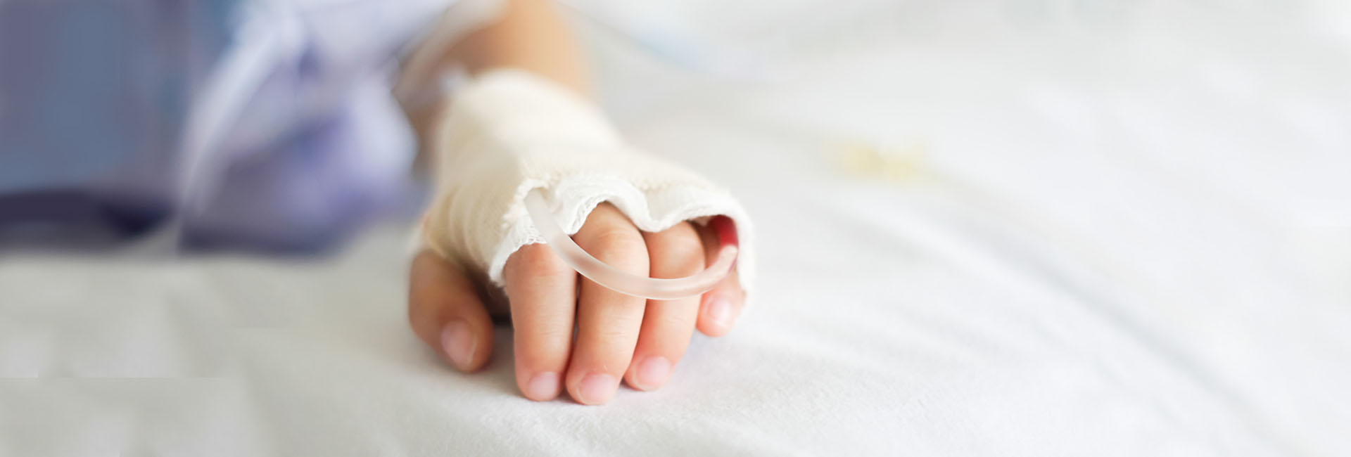
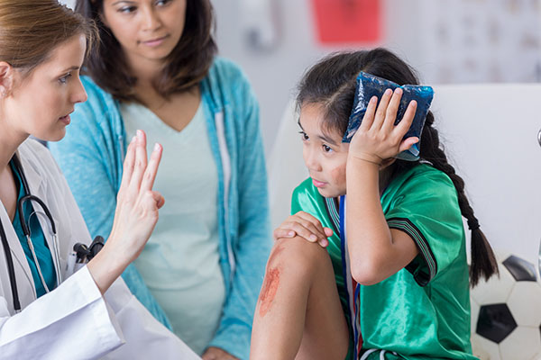
Approximately 100,000 children end up in the hospital every year for head and brain injuries due to falls, sports injuries, bike accidents, car accidents, child abuse, and more. Medical professionals used to believe that children suffering from brain injuries were more resilient in recovery because their brains would “rewire” over time. However, mounting evidence seems to indicate that children could be even more susceptible than adults to permanent brain damage. Therefore, it’s important to have your child examined as quickly as possible after a head injury occurs, even if your child appears to be fine.
Types of brain injuries we treat include:
- Skull fractures – Although these types of injuries typically heal on their own, they need to be observed and monitored.
- Hemorrhages – Also known as hematomas, hemorrhages may require surgery, depending on where the bleeding occurs.
- Traumatic brain injuries – These are the result of a blow to the head and may not be apparent immediately. There are several types of TBIs, beginning with concussions and going to the more severe kinds of injuries that cause permanent brain damage.
The treatment we recommend for your child depends on the type of brain injury s/he has. Options could include:
- Surgery to repair breaks and/or bleeding
- Drain or shunt placement to reduce pressure
- Prescribing medications to prevent and/or control seizures, muscle spasms, blood clots
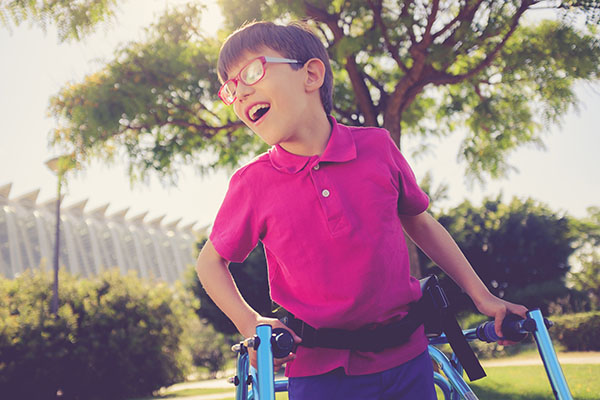
Cerebral Palsy
Cerebral palsy is the term used to describe a group of neurological disorders that affect the part of the brain that controls muscle movement. It’s the leading cause of childhood disabilities, affecting 3.3 children per 1,000. Cerebral palsy is caused by damage or abnormalities to a child’s developing brain, which, in turn, disrupts the brain’s ability to control movement and maintain posture and balance.
Children with cerebral palsy typically begin displaying symptoms in infancy or early childhood. These symptoms include:
- Weakness in one or more extremities
- Tremors or random involuntary movements
- Difficulty with movements like tying shoes or buttoning shirts
- Delays in reaching motor skill milestones
- Lack of general muscle coordination
- Spastic movements or exaggerated reflexes
- Crouched or “scissored” gait
- Walking on toes
- Excessive drooling
- Difficulty swallowing or speaking
Cerebral palsy doesn’t always cause extreme disabilities – it will vary from one child to another. Although there is no cure for cerebral palsy, a child’s capabilities can improve through a combination of medication and therapies.
Some types of surgery can also improve symptoms of cerebral palsy, especially for children who suffer from severe spasticity and stiffness that make walking and moving difficult or painful.
At Brain & Spine Surgeons of New York, we are experts in the surgical procedures designed to help children with cerebral palsy live healthier lives.
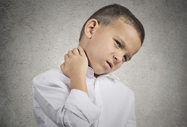
Chiari Malformation
A Chiari malformation is a deformity of the brain that affects the lower part, which is called the cerebellum. The cerebellum is the vital part of the brain responsible for controlling muscle movement and coordination. Chiari malformation causes the child’s cerebellum to be pushed into the space normally occupied by the spinal cord.
The majority of children who suffer from Chiari malformation are born with it, but some develop the condition as they mature and grow. A few of the most common symptoms associated with Chiari malformation include:
- Problems with balance
- Headaches in the rear of the head
- Numbness and weakness
- Neck pain
- Tingling or pain in their arms
- Uncontrollable eye movements – typically from side to side
- Decreased gag reflex or problems swallowing
- Sleep problems caused by sleep apnea
- Issues with certain motor tasks, such as cutting with scissors or writing
Many children with Chiari malformation do not experience symptoms and may never require medical treatment. Even so, children diagnosed with this disease should continue to visit their physician for regular checkups. These checkups will ensure the child’s medical condition isn’t changing.
If symptoms are present, treatment is designed to alleviate the symptoms and prevent any further spinal cord and brain damage. Physicians commonly recommend medicines for neck pain, headaches, and other symptoms. The doctor may also suggest minimizing any activity that worsens the symptoms.
However, those with more severe symptoms should be treated by a neurosurgeon to determine whether surgery is appropriate. The most common Chiari malformation surgical procedure is decompression. Decompression is designed to restore the flow of cerebrospinal fluid, relieve the pressure on the spinal cord, relieve the pressure on the brain, and prevent damage to the spine and brain. The procedure also includes measures to reduce spinal pressure in the area.

Craniocervical Abnormalities
Craniocervical abnormalities are essentially cervical spine defects in the highly complex area where the upper cervical spine and skull join together. These abnormalities can squeeze and damage the spinal cord nerves, which lead directly to the brainstem and brain. Craniocervical abnormalities may weaken the spine and cause serious neurological disorders. Symptoms of this condition include:
- A burning sensation that spreads down legs, buttocks, or arms.
- Weakness, numbness, or cramping in hands
- Numbness in feet
- Back stiffness and pain
- Ear ringing and pain
- Involuntary movement of the eyes
- Difficulty with leg and hand coordination
- Off-balance feeling
While craniocervical abnormalities may be present at birth, they may also develop later in life as a result of disorders or injuries.
Craniocervical abnormalities must be diagnosed through a CT scan or MRI. After diagnosis, the treatment typically involves reduction then stabilization with an external device or through surgery. Surgical treatments may include:
- A surgical procedure to stabilize the spine
- Surgical decompression to help alleviate the pressure on the spine and brain
- The Expanded Endonasal Approach (EEA) is a minimally invasive procedure where the surgeon accesses the skull through the nasal cavity. This process results in no incisions, faster recovery, and a shorter hospital stay in comparison to traditional brain surgery

Craniofacial Syndromes
Craniofacial syndromes refer to a pediatric condition where a child has one or more abnormalities of the face and/or the head. This condition can happen when the soft plates of a baby’s skull close too soon, or in an unusual way, typically resulting in disfigurement or unusual facial appearances. Craniofacial syndromes can also result from other abnormal growth patterns of the face or skull, disease, or trauma.
There are numerous different craniofacial syndromes, the most common of which include Crouzon, Apert, Pfeiffer, Muenke, and Saethre-Chotzen syndromes. Each syndrome has a different set of potential complications, requiring a unique approach to surgical management. The expert pediatric neurosurgeons at BSSNY have knowledge and experience in treating even the most challenging craniofacial abnormalities.
After examination and imaging, we may recommend surgery to correct the physical formation of the cranial and facial bones and maximize the function for the child. Often the pediatric neurosurgeon will perform the surgery in conjunction with a pediatric craniofacial plastic surgeon.
BSSNY’s pediatric neurosurgeons are fellowship-trained and fluent in endoscopic approaches to these kinds of restoration surgeries, which can provide a simpler technique that may minimize blood loss, complications, and recovery time. With these types of endoscopic procedures, there is typically minimal scarring.

Craniosynostosis
A newborn baby’s skull is made up of seven separate bones that haven’t yet fused together. Normally, fusion happens gradually and isn’t completely fused until the late teen years. However, craniosynostosis is a birth defect where two or more of those bones close prematurely before the baby’s brain is fully formed.
A baby’s brain grows quickly over the first two years of life, so it’s important that the bones remain open. So as the brain growth continues, the heads of infants with craniosynostosis take on a misshapen appearance. Left untreated, the condition can interfere with normal growth of the brain and skull.
Generally speaking, the signs of craniosynostosis are typically noticeable at birth, but they’ll become more apparent during the first few months of a baby’s life. Look for:
- A misshapen skull
- Disappearance of the fontanel on your baby’s skull
- Development of a raised, hard ridge along affected sutures (where the bones fuse together)
- Slow or no growth of the head as your baby grows
Treatment of craniosynostosis almost always involves surgery to correct the shape of the head and allow for normal brain growth. Surgical treatment allows your baby’s brain adequate space to grow and develop.
Although neurological damage can occur, most children have normal cognitive development and achieve good cosmetic results after surgery. Early diagnosis and treatment are key.

Epilepsy
Epilepsy is a condition that affects the child’s central nervous system and deals with electrical signals of the brain misfiring. As a result of these electrical disruptions or misfirings, the nerve cells experience temporary problems with communication, which causes seizures.
Seizures can be considered to be “electrical storms” that makes the brain react to things in unintended ways. If a child has one seizure – or even several seizures – it doesn’t necessarily mean the child suffers from epilepsy. Children who suffer from epilepsy are more likely to have several seizures over long periods of times, such as months or years.
While approximately 3 million Americans suffer from epilepsy, most of the newly diagnosed cases are in children. Anyone can get epilepsy at any age, and some kids outgrow their seizures by their teens. It’s important to understand that epilepsy is not a mental illness and isn’t the only source of childhood seizures. This condition isn’t contagious and has no direct relation to a child’s intelligence.
The physician will use various tests and exams to arrive at the optimum treatment plan for epilepsy. In most cases, the first line of defense for seizure prevention and epilepsy management is medication. Many kids are treated successfully with a single medication.
However, if the first medicine doesn’t work, the doctor may try a second or third medication before they decide to use a combination of medication. With epilepsy, no medication is the end-all for the condition, and side effects are a possibility. The most common side effects include:
- Behavioral or mood concerns
- Tiredness
- Decreased alertness
Head, Neck, and Skull Base Disorders
At Brain & Spine Surgeons of New York, we provide comprehensive care to children who have head, neck and skull base disorders, including congenital anomalies and tumors. Our board-certified pediatric neurosurgeons will give your child expert, compassionate care using the most advanced surgical techniques.
We understand the complexity of the region, so our treatment approach focuses on innovative minimally invasive, endoscopic technologies, which maximize outcomes while reducing postoperative effects of surgery. Where possible, we perform the surgery through the natural orifices of the face to minimize scarring. The result is less blood loss, faster recovery, and excellent outcomes.

Hydrocephalus
According to the National Institute of Neurological Disorders and Stroke (NINDS), anywhere from one to two babies out of every 1,000 are born with hydrocephalus. Hydrocephalus is a condition that occurs when cerebrospinal fluid (CSF) fluid doesn’t properly drain from the brain. CSF is the watery, clear fluid that cushions and surrounds the brain and spinal cord.
When the cerebrospinal fluid fails to drain, it places the brain under excessive pressure and causes swelling, which damages the brain tissue. Hydrocephalus is directly related to intellectual, developmental, and physical impairments.
Hydrocephalus can begin before the baby is born from spinal column birth defects and genetic abnormalities. It can also begin while the child is in the womb as a result of certain pregnancy infections, such as rubella. Hydrocephalus can also occur in infants, toddlers, and adolescents due to:
- Central nervous system tumors
- Head trauma
- Injuries before, during, or after delivery
- Brain bleeding during or after delivery
- Meningitis and other central nervous system infections
If left untreated, hydrocephalus can be fatal. While treatment may not reverse the brain damage that has already happened, the goal of treatment is to prevent the brain damage from progressing further. Physicians typically explore one of the following medical treatment options:
- Shunt Insertion – A shunt, which is a drainage system comprised of a valve and a long tube, is surgically inserted. The valve helps facilitate the flow of cerebrospinal fluid in the right direction at a normal rate. Excess fluid from the brain drains out on the other end of the tube into the abdominal cavity or chest.
- Endoscopic Third Ventriculostomy (ETV) – As an alternative to the shunt procedure, a ventriculostomy involves creating a hole between or at the bottom of the ventricles. The placement allows the cerebrospinal fluid to safely leave the brain. This process involves the use of a tiny endoscope, which is a camera, to complete the procedure. Children who have an ETV may avoid the need to have the shunt replaced, getting infections, and other complications associated with shunts.

Moyamoya Disease
Moyamoya disease or Moyamoya syndrome is a very rare, but extremely serious pediatric condition. Children who suffer from this disease have narrowed and thickened internal carotid arteries. The carotid arteries are responsible for supplying blood to the important areas of the brain.
Because these arteries are thickened and narrowed, the natural flow of oxygen-rich blood to the child’s developing brain begins to slow down gradually. The constriction also increases the probability of blood clot formation.
The initial symptom of Moyamoya disease is typically a stroke or a mini-stroke, which is medically referred to as recurrent transient ischemic attacks. These symptoms may be accompanied by seizures or paralysis and muscle weakening on one side of the body. Children may also experience cognitive decline and headaches.
One of the most common medical recommendations for Moyamoya disease is aspirin, which may be an effective way to bolster blood flow to the brain. However, most children who suffer from Moyamoya syndrome will require surgery to restore blood supply to the brain.
The encephalo-duro-arterio-synangiosis (EDAS) treatment is an indirect bypass method of revascularization. During EDAS, a temporal superficial artery is connected to the surface of the brain. A hole is then created in the skull beneath the artery, which is sutured to the brain surface. Over time, the process of angiogenesis forms small, arterial vessels to the brain. It usually takes anywhere from six to eight weeks after the procedure for the new blood supply to form.

Pediatric Brain Tumors
Pediatric brain tumors are masses of abnormal cells located in a child’s brain or the tissue and structures nearby. There are many types of pediatric brain tumors – some of which are benign, while others are cancerous.
Brain tumors in children typically begin when normal cells have DNA mutations, which grow and divide at increased rates. This results in a mass of abnormal cells that ultimately forms a tumor.
Common forms of pediatric brain tumors include:
- Choroid plexus carcinoma – a rare cancerous brain tumor that most often occurs in children under two years old.
- Craniopharyngioma – a noncancerous brain tumor that begins near the brain’s pituitary gland and secretes hormones that affect the function of the pituitary bland.
- Embryonal tumors – a cancerous brain tumor that starts in the fetal (embryonic) cells in the brain. Occurs mainly in babies and young children.
- Ependymoma – a malignant tumor that forms in the tissues of the brain and spinal cord.
- Glioma – one of the most common of brain tumors, gliomas can affect brain function and can be life-threatening.
- Medulloblastoma – this is a cancerous brain tumor that starts in the cerebellum and can affect balance, motor function, and movement.
- Pineoblastoma – this is a rare, aggressive type of cancer that begins in the cells of the brain’s pineal gland, which controls the sleep-wake cycle.
As you can imagine, treatment for pediatric brain tumors can be quite different from treatment for adult brain tumors, so it’s very important that you enlist the help of an experienced pediatric neurosurgeon.
Your child’s prognosis and our approach to treatment will depend on the type of tumor your child has, its location, and whether it has spread. Other important factors include your child’s age and general health.
Pediatric Spinal Cord Tumors
Pediatric spinal cord tumors are a fairly common type of childhood cancer in which abnormal cells form in the tissue inside the spinal column. Although these tumors can be cancerous or non-cancerous, they should be treated immediately because they can cause serious health problems as they grow and place pressure on the spinal cord.
Nerves located within the spinal cord connect the brain to other parts of the body, carrying messages that make the body move, taste, feel, etc. When a tumor develops, body functions may be affected, such as:
- Partial paralysis and/or trouble walking
- Spinal deformity
- Back pain or pain that spreads from the back towards the arms or legs
- A change in bowel habits or trouble urinating
- Leg weakness
In addition, some children aren’t able to reach certain growth and developmental milestones such as sitting up and walking.
Children and teens with brain and spinal cord tumors and their families have special needs. At BSSNY, we work closely with the child’s primary care doctor in order to maximize a successful outcome.
Typically, we will perform surgery to remove or reduce the size of the tumor and alleviate the pressure on the spinal column caused by the tumor. Once the surgery has been performed, a pathologist examines tumor tissue to determine the exact type of tumor. Our team is led by one of our board-certified pediatric neurosurgeons who is an expert in the treatment of pediatric spinal cord tumors.

Spina Bifida
Spina bifida is one of the several types of spinal dysraphism, which is a term used to explain malformations of the spinal cord. As one of the most common birth defects in the world, spina bifida occurs when the embryo’s developing neural tube fails to close correctly. The neural tube is the precursor to the spinal cord and brain.
Spina bifida may affect the spinal cord coverings, the spinal cord itself, and/or any of the bones of the spine. This condition can cause serious infections, problems with bowel and bladder function, paralysis, and hydrocephalus.
In the most severe form of spina bifida (open spina bifida), an open channel exists from the skin to the spinal cord. This defect is usually visible to the naked eye in a newborn or can be detected through fetal ultrasounds. As a result, this form of spina bifida is always diagnosed in newborn infants or in utero.

Tethered Spinal Cord Syndrome
Tethered spinal cord syndrome is a neurological condition caused by the attachment of tissue that restricts the spinal cord movement inside of the spinal column. Because of these attachments, the spinal cord is stretched abnormally. The tethered spinal cord syndrome is closely related to spina bifida.
It’s estimated that anywhere from 20 to 50 percent of children born with spina bifida defects that were repaired shortly after they were born will require a procedure to untether the spinal cord. This is because the spinal cord remains in the same location after the spina bifida procedure. As the child continues to grow and mature, the spinal cord may become stretched, which interferes with the spinal cord’s blood supply and causes damage.
Children suffering from tethered spinal cord syndrome may experience difficulties walking or standing, back pain, leg pain, urinary and/or fecal incontinence, and numbness or weakness in the feet or legs. Causes of tethered cord syndrome include:
- Myelomeningocele
- Spinal Lipoma
- Dermal sinus tract
- Fatty filum
The recommended treatment for “untethering” the spinal cord is surgery, which may also reverse or prevent progressive neurological symptoms. However, the procedure is only performed if there are symptoms of deterioration or clinical signs.
The type of surgery depends on the actual cause of the tethered spinal cord syndrome, such as myelomeningocele, dermal sinus, Spinal Lipoma, or fatty filum. Regardless of the cause, prompt surgical intervention is typically the best solution. Most children can return to their normal activities after a few weeks after the procedure has been successfully performed.

Vascular Malformations of the Brain
Vascular malformations of the brain is a general term used to classify conditions that affect the blood vessels in the brain. During the fetal development stage, abnormal blood clusters form and can have serious repercussions if left untreated. Vascular malformations may cause seizures, headaches, bleeding in the brain, and strokes.
These conditions include:
- Cavernomas or cavernous malformations of the brain are vascular abnormalities of the central nervous system. This condition is characterized by a cluster of dilated, abnormal vessels.
- Cerebral aneurysms explain the ballooning, bulging, or dilation of part of the artery or wall of a vein in the brain.
- Arteriovenous Malformations (AVM) are tangles of abnormal blood vessels that connect veins and arteries to the brain. These veins are tasked with carrying oxygen-depleted blood back to the heart and lungs. The arteries are responsible for transferring oxygen-infused blood to the brain and heart.
Vascular malformations will typically grow at a rate proportionate to the child’s development. Treatment options will vary based on the location and severity of the malformation. While advancements in medicine are continually introducing new solutions, the current treatment options include:
- Resection or surgical removal of the vascular malformation of the brain.
- Multiple embolization is a procedure where pellets are placed in the circulatory system to constrict blood flow from and to the abnormal blood vessels.
- Irradiation or radiation treatment using hyper-focused lasers to minimize the vascular malformation.